What is the Neck Capsule?
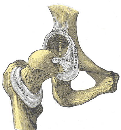
Capsule of hip joint
The articular capsule (capsular ligament) is strong and dense.
Above, it is attached to the margin of the acetabulum 5 to 6 mm. beyond the glenoidal labrum behind; but in front, it is attached to the outer margin of the labrum, and, opposite to the notch where the margin of the cavity is deficient, it is connected to the transverse ligament, and by a few fibers to the edge of the obturator foramen.

Photo, left: Capsule of hip-joint (distended). Posterior aspect
Photo Right: Hip-joint, front view. The capsular ligament has been largely removed. (Capsular ligamnet visible at center.)
It surrounds the neck of the femur, and is attached, in front, to the intertrochanteric line; above, to the base of the neck; behind, to the neck, about 1.25 cm. above the intertrochanteric crest; below, to the lower part of the neck, close to the lesser trochanter.
From its femoral attachment some of the fibers are reflected upward along the neck as longitudinal bands, termed retinacula.
The capsule is much thicker at the upper and forepart of the joint, where the greatest amount of resistance is required; behind and below, it is thin and loose.
It consists of two sets of fibers, circular and longitudinal.
The circular fibers, zona orbicularis, are most abundant at the lower and back part of the capsule, and form a sling or collar around the neck of the femur.
Anteriorly they blend with the deep surface of the iliofemoral ligament, and gain an attachment to the anterior inferior iliac spine.
The longitudinal fibers are greatest in amount at the upper and front part of the capsule, where they are reinforced by distinct bands, or accessory ligaments, of which the most important is the iliofemoral ligament.
The other accessory bands are known as the pubocapsular and the ischiocapsular ligaments.
The external surface of the capsule is rough, covered by numerous muscles, and separated in front from the Psoas major and Iliacus by a bursa, which not infrequently communicates by a circular aperture with the cavity of the joint.
This article was originally based on an entry from a public domain edition of Gray's Anatomy. As such, some of the information contained herein may be outdated. Please edit the article if this is the case, and feel free to remove this notice when it is no longer relevant.
Updated December 2008