False Profile X-Rays
Anatomical and radiological correlation of Lequesne's "false profile"
Abstract
Lequesne introduced a radiological projection, which is an oblique view of the edge of the acetabulum, to diagnose arthrosis affecting the anterior part of the joint and to measure the anterior coverage of the femoral head. In this study, we attempted to determine the anatomical correlation of his technique. Fifteen in vitro hemipelvises underwent radiography according to Lequesne's description, using metallic markers and wires to mark physical landmarks. According to geometric laws, the points used by Lequesne do not correlate anatomically. Although Lequesne's technique allows a diagnosis of acetabular dysplasia, measurements are on average 5.5° less than those made anatomically. The French version of this article is available in the form of electronic supplementary material and can be obtained by using the Springer LINK server located at http://dx.doi.org/10.1007/s00276-002-0038-1.
False Profile View
Lequesne and de Seze view
- Helpful for: DDH, (Developmental Dysplasia in Hips)
- Evaluate: anterior coverage of the femoral head
- Measeure ventral inclination angle. Normal >25º.
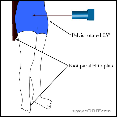
Position:
Patient standing with the affected hip on the cassette, the ipsilateral
foot parallel to the cassette and the pelvis rotated 65° from the plane
of the cassette.
Beam: centered on the femoral head perpendicular to the cassette
Reference: Garbuz DS, CORR 2004;418:18, Lequense M, Rev Rhum Mal Osteoartic 1961;28:643