How to Email X-Rays
X-Rays in standard film format
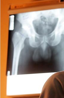
You can take a digital camera with you to the doctor’s office and just snap some pictures while the x-ray is up on the light box. Take several to make sure you get at least one or two clear shots. Then make sure the size of the file is not too large to email (see below for instructions to compress file).
You can also take digital pictures of your X-Ray film by holding the X-Rays up to a white background like a blank Word document. Or place it on top of a glass coffee table with a white background. Sometimes a window will work if there is a clear background.
To compress your x-ray once it is in jpeg format read below. DO NOT SEND ZIPPED FILES, AGAIN A ZIP FILE WILL NOT WORK, IT MUST be compressed this way as instructed below. Please include your AGE, Height and weight, any health conditions such as liver or kidney disorders, diabetes, etc. Any heart or breathing problems, anything that might affect surgery, meds you are taking, activity level prior to hip going bad and detailed symptoms.
Once you have your x-rays in jpeg format and have them on your computer, you will need to compress the files prior to emailing the x-rays. If you open them up using Microsoft Office Picture Manager, you will see a screen like this
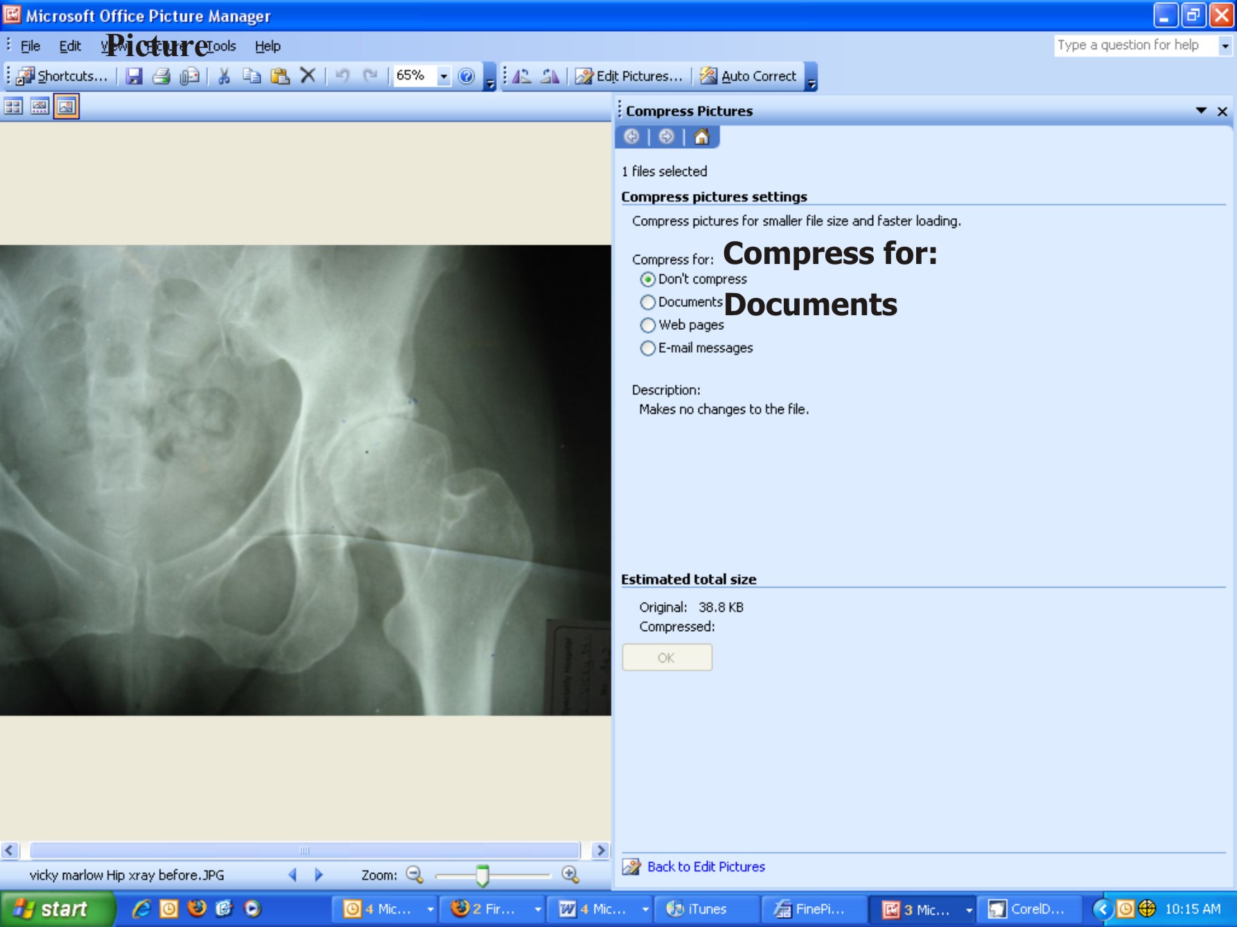 See on the top I have bolded the menu item Picture,
select that and you will see a drop down menu, click on Compress.
After you do that you will see this screen to the left and a choice of
See on the top I have bolded the menu item Picture,
select that and you will see a drop down menu, click on Compress.
After you do that you will see this screen to the left and a choice of
Compress for:
O Don't Compress
O Documents
O Web pages
O Email messages
Select Documents
Then SAVE the file. Name the file with your name, or initials and the date your x-ray was taken, that way if you are sending several taken at different times, they will be easily recognizable by date they were taken. Mine might look like Vicky120106 or VM120106.
DCM X-Ray images
Thanks to Ron van Mierlo for providing the majority of info below
For Ron’s info: http://www.resurfacingscan.be/eindex.htm select 'English', 'Technical' and 'DCM X-Ray images'
Intro
DCM is a digital format that is commonly used in the medical world today. The letters DCM are taken from the abbreviation DICOM that in turn stands for ” Digital Imaging and Communications in Medicine”. The images with the extension DCM can originate from CT-Scan, MRI and other devices. With DICOM now accepted as the medical standard, image data can easily be exchanged between hospitals, specialists, universities etc. through networks, the Internet and e-mail, it is even possible to show life images somewhere else in the world.
To convert from CD to JPEG
As hip patients you may receive your X-Rays on a CD with the images in DCM format which will be found on the disk. Usually only X-Rays are needed, but in certain complicated cases, other tests may be required, example a bone density scan for women above a certain age or sometimes an MRI.
Images from the performed test(s) are stored at hospitals or radiology
departments, but can be requested in the form of one or more CD-disks
(with different types of tests performed, there may be one CD for each
test). You will be required to sign a release form in person with the
HIPAA laws. Contact your doctors office or radiology department and
arrange a time to pick them up and tell them you will be there in
person and will sign the release form for them. They are your right to
have, so do not allow them to tell you they need a doctor to order
them. The X-Rays should be recent preferably within 90 days, six
months the latest.
Sometimes they may charge for the CD, I paid $20 U.S., it will vary.
Viewer
A”Viewer program” comes with the CD-disk but which exact program it is
and which options the viewer offers differs from hospital to hospital,
sadly enough, there does not appear to be any common viewer. The viewer
program normally starts automatically after inserting the CD into your
computer and it begins to show your images. The images can be very
large, somewhere between 2Mb or 20MB per image.
Depending on the viewer program in use, an option may exist for the
”export” of images, while in other cases the option to “save as” may
exist, or the DCM images can be saved in another image format that is
supported by the viewer program, like for instance BMP, JPG, GIF or
TIF. The advantage for you as a patient is that the created file can be
much less in size, up to one tenth of the DCM image and will be more
acceptable to most computer programs, that is not so with the DCM
images. If you have the choice for the size to export, then 150kb –
300kb is what you should select. It would be impossible for the
surgeons to receive e-mails from patients with large size images and
since many surgeons receive a lot of these, it is best not to lock up
their systems with overload. There is still enough detail left in JPG
images of 150kb and in some cases if that is not good enough, then the
surgeon will let you know what he/she requires.
Creating JPG images from DCM X-Rays
When the CD’s viewer program doesn’t offer any means to ”export to” or
”save as” JPG images then there are several other ways to create your
JPG images.
1.) Both Macs and PCs offer ways to create so-called screendumps, useable for our purpose.
Mac:
1a.) With the viewer program running and the requested X-Ray
image shown screen-filled you can with the key combination
”Shift-Apple-3” (press and hold ’Shift’, then ’Apple’ and finally ’3’)
create a useable image, that normally will finish up on the desktop.
The same key combination can be repeated for other X-Ray images shown
in the viewer. When you even would like to have the option to crop
images and cut away some parts then you can use the combination
”Shift-Apple-4”.
1b.) If you intend to do more with DCM images apart from just
conversions a program called “OsiriX” used, it can be downloaded from
the Internet.
PC:
1a.) With the viewer program running and the requested X-Ray 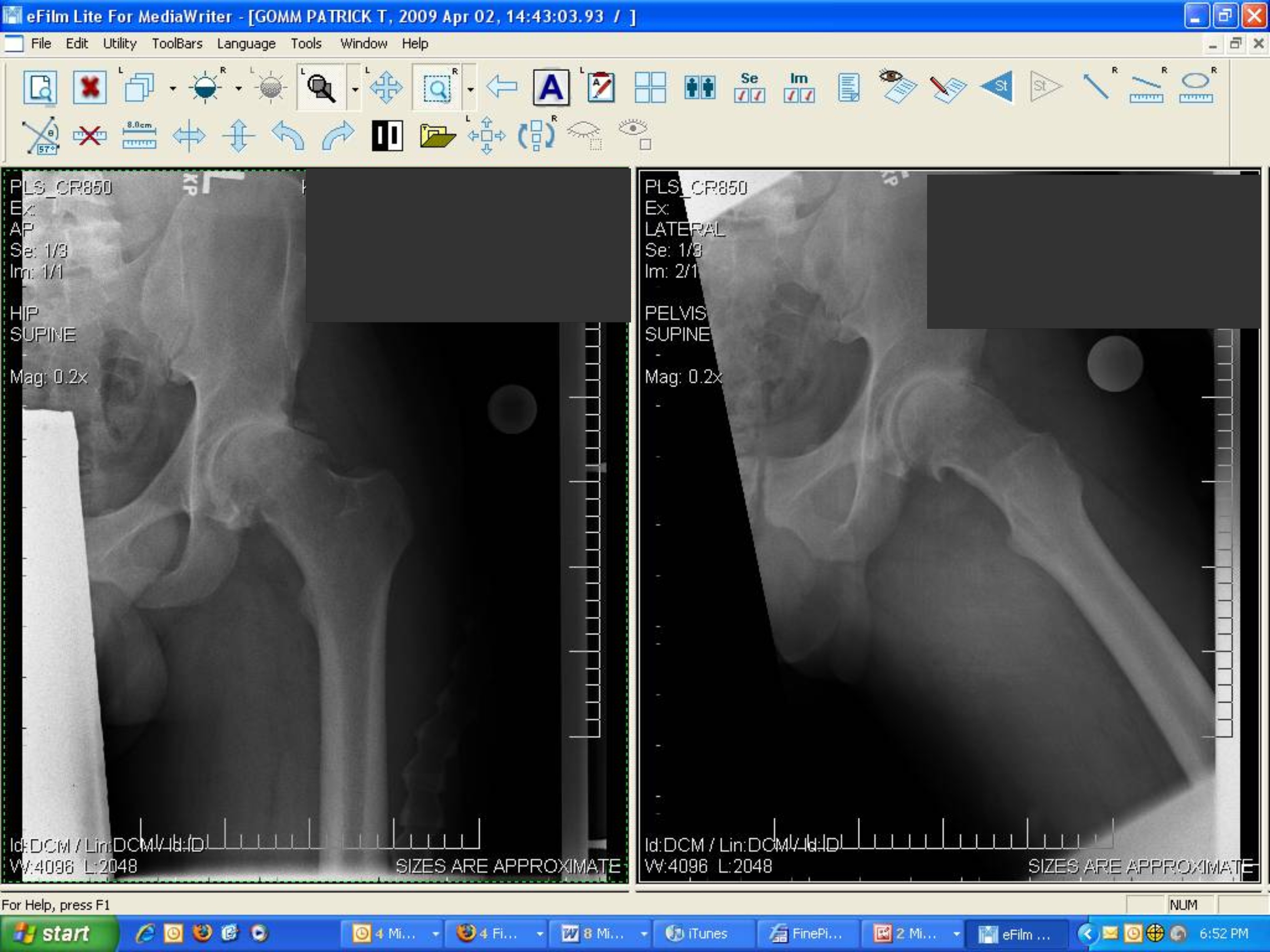 image shown screen-filled you can press on the
”PrtSc” (Print Screen) button and a screen image is created
and kept in RAM memory. After opening the little standard program
”Paint” that comes with all Windows based systems, you can now choose
“Edit” from the menu row, followed by “paste” to see the image appear.
Thereafter you will have to save this image by selecting ”Archive” and
”Save as”…there you will have to select in the file format window the
alternative ”JPEG (*.JPG,*.JPEG,*.JPE,*.JFIF)”.
For each new image you’ll have to repeat the procedure from point 1a.
1b.) It is possible to download a powerful little program called ”Irfanview”.
http://www.irfanview.com/
It has the capability to show still images of most digital formats and
even some video formats. From the website of this program you can also
download plugins, like in our case components for the support of DCM
eller DICOM images. With all that installed on the computer you can
show and manipulate your DCM images and save them in every possible
digital format and size.
Finding the images on the CD
With the computer’s ”Search” function that is found right after
clicking on the ”Start” button in Windows based systems, you can search
for the images with the DCM extension. With “Explorer” you can of
course browse around and look for a folder on the CD that holds the DCM
images. Some CD-disks hold the images in a folder named ”DICOM”, this
would be simple but in reality any odd folder name can be used.
The extension DCM can in some cases miss completely behind the file
names. So in those cases you are best off looking for some extra large
files that most likely are placed in the root directory or main folder
of the disk and have file names that contain some data like your name,
the date the images were taken, date of birth, social security number
etc. Those files will then have to be copied to a place on your hard
disk where each of them have to be given their own extension DCM (or
dcm). Therafter it will be possible again to open up those files with
programs that recognize the DCM format.
There may be some cases where a patient’s diagnosis can not be done
from X-Rays alone. If that happens, the surgeons will reply and
indicate which specific additional tests and information are required.
If you are still having trouble converting your x-rays, you can email Tom at
v_5whiteth@ yahoo.com, just delete the space or This e-mail address is being protected from spam bots, you need JavaScript enabled to view it .